Advantages for the Physician
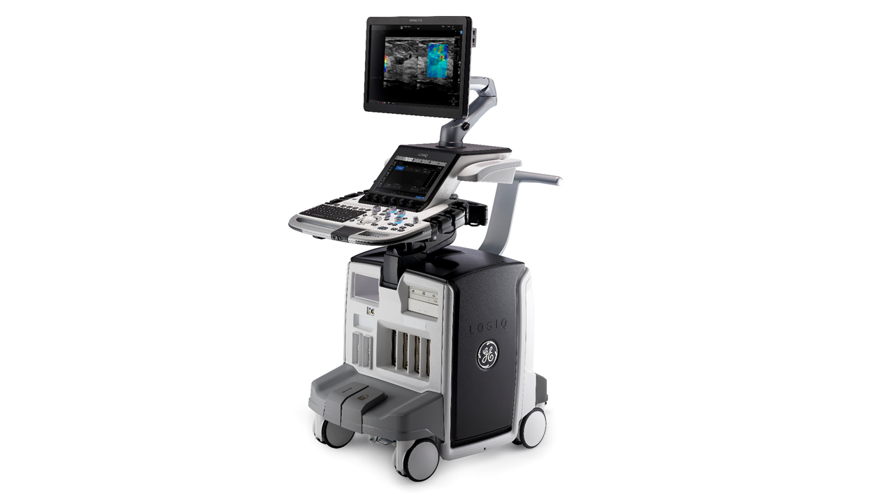
The LOGIQ™ Ε10 ultrasound system literally redefines every element of the diagnostic chain, from the pulse of the sensors to the purity of the pixels. The result is the highest level of diagnostic performance GE Healthcare has ever offered – a leap forward that one must see in order to believe.
Every day, you have the opportunity to make a real difference in the lives of your patients. At GE Healthcare, we want to allow you to do this with the new top-of-the-line LOGIQ ™ E10 model - backing up your experience and helping you take your practice to the next level. The new LOGIQ E10 is designed for you - so you can make a reliable and effective diagnosis using integrated tools and achieve a new summary workflow standard
- Impressive image quality & excellent clinical versatility to respond to a wide range of examinations, including abdominal, vascular, obstetric, gynecological, neonatal, pediatric, urological, intracranial, cardiac, musculoskeletal, interventional and minor applications.
- Easy workflow with a user-friendly console, “onboard” automation and productivity packages that help speed up the workload of very busy programs.
- Special tools respond to complex cases, in which information obtained through technologies, such as Elastography can support fast, accurate estimates.
Impressive image quality & excellent clinical versatility for knowledge expansion
The results of these imaging developments are improved speed and reliability. The LOGIQ Ε10 system contributes to the rapid acquisition of the necessary information, both in everyday and in difficult cases, in order to support decisions that promote clinical reliability.
“Smart” acoustic architecture – Automatically adapts to the patient’s body habits
XDclear processing system – Active “background” mode that optimizes image quality
High definition, wide screen display – Features state-of-the-art OLED display resulting in:
- Maximum Contrast resolution and tracing capacity
- Increased image quality regardless of the physician’s point of view
- Increased resolution regardless of the brightness of the examination area
Specialized tools for complex questions
B-Flow Imaging – Leaving doppler limitations behind
Through direct, real-time visualization of blood flow echoes, the updates of B-Flow™ / B-Flow Color imaging optimize hemodynamic flow assessment in a wide variety of studies to better evaluate micro- and macro-vascular structures, areas suspicious of malignancy, thrombosis, vascular stenosis inflammations, etc. The technique is particularly useful for the study and identification of areas suspicious of malignancy, which present impaired hemodynamic behavior and are impossible to detect with conventional angiography techniques, such as for example the use of Doppler.
Elastography – More information for patient management
Strain Elastography – A powerful tool that includes mapping, estimating the pressure (strain) or the deformation of the tissue in the area of interest after applying pressure. It can be applied to a wide range of tests, anatomical areas and organs such as abdomen, liver, superficial organs (breast, thyroid gland), study of musculoskeletal diseases, intra-abdominal organs (prostate, female reproductive organs) for the assessment of the findings obtained.
Shear Wave Elastography – By allowing non-invasive two-dimensional quantitative assessment of tissue hardness, this tool can prove to be of particular value for assessing soft tissue status. The technique is applied to multiple anatomical areas and organs of the body (deep and superficial), to evaluate the presented findings. Quantitative data are exported in kPa units (elasticity) & m/s units (speed in meters per second), to increase the diagnostic capacity (sensitivity, specificity, etc.) for growth characterization.
Compare Assistant – seeing the past in real time
The Compare Assistant feature allows physicians to have easy access to a previous study – an ultrasound or a study using other means and imaging, side-by-side in real-time with up-to-date images of the patient, helping to enhance the reliability and efficiency of the examination.
Color Flow Quantification - quantitative estimation of color flow.
It enables quantification of tumor vasculature, areas suspicious of malignancy or inflammation of musculoskeletal structures (e.g., rheumatoid arthritis). The results obtained are used both in clinical investigations and for the creation of treatment planning and monitoring protocols. Follow-up of treatment in patients using the Flow Quantification technique is of great importance especially for cases of oncology patients, where rapid results and redesign of treatments are required.
Coded Contrast Imaging A technique for detecting and displaying the acoustic signals of the fundamental 2nd harmonic frequency derived from the injection of contrast materials, of a suitable mechanical index. The technique uses a large number of all types of probes, such as Convex/ Μicroconvex, Sector Phased Array & Linear, covering all possible applications used in ultrasonography and require study with the use of a contrast medium, for perfusion studies in a variety of organs and the in-depth study of examinations performed for example in the upper-lower abdomen, superficial organs (breast, thyroid gland, salivary glands, etc.), intra-abdominal organs (prostate, etc.).
It enables Quantitative data to be exported via graphic representations (Time Intensity Curve (TIC) Analysis.
Volume Navigation: Features state-of-the-art synchronized ultrasound imaging technique in real-time with CT, MRI, PET/CT, SPECT scans/sequences and previous ultrasonography examinations allowing volumetric navigation in these sequences as well as with a combination of contrast enhanced techniques and elastography.
Enables Volume Navigation of the following examinations:
- upper & lower abdomen as well as for pediatric examinations (very important Follow up to avoid radiation)
- prostate, female reproductive organ examinations
- vascular examinations, superficial organ examinations, musculoskeletal examinations
It enables the performance of precision guided biopsy Real time Needle Tracking in a wide variety of probes that support the Volume Navigation GPS Technology feature, in multiple organs, such as breast, liver, prostate, etc.
This techniques allows the course of needle to be pre-planned before it even enters the patient’s body, thus ensuring not only the safe passage of the needle through the anatomical structures (such as nerves, vessels, etc.) but also the accuracy of sampling from the area of interest. The technique has a wide application in the calculation - counting of multifocal lesions in different anatomical areas, thus avoiding the possibility of counting the same lesion more than once.
High performance probes for everyday and specialized applications
The LOGIQ Ε10 system offers a wide selection of probes from the E-Series and XDclear probe series to support a wide variety of applications. The revolutionary XDclear probe technology delivers extremely high resolution trace capacity at impressively high depths for imaging even the most difficult, obese and of larger physique patients.
C1-6-D: XDclear state-of-the-art probe that provides excellent in-depth resolution for excellent clarity on abdominal organ examinations
L2-9-D: linear probe that supports specialized imaging with large foot print and field of exceptional depth for imaging of the peripheral and deep vessels and carotids.
ML6-15-D: matrix array linear probe of advanced technology that provides high resolution imaging in the structural details of small parts, neonatal, pediatric, musculoskeletal and peripheral vascular applications.
IC5-9D probe for high quality intravaginal and intrarectal examinations
Advantages for the patient
Henry Dunant Hospital Center now features the most modern high-tech General Electric ultrasound scanner, LOGIQ E10.
It provides high definition images for the following examinations:
- upper-lower abdominal organs (liver, bile, spleen, kidneys, renal arteries, bladder)
- internal genitals (uterus, cervix, ovaries, prostate)
- breast, thyroid gland, glands, scrotum
- musculoskeletal system
- superficial and deep veins, carotids, aorta, upper/ lower extremity vessels
- examinations with the use of contrast medium
- elastography examinations (strain & shear wave)