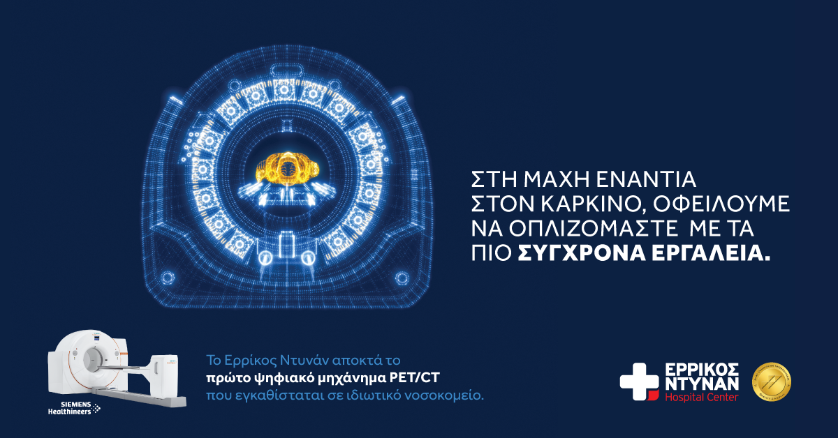
With the introduction of the latest generation equipment, specifically the first digital PET/CT scanner operating in private Greek hospitals, the Henry Dunant Hospital Center establishes itself as a model Diagnostic and Therapeutic Nuclear Medicine Center. This development provides diagnoses of exceptional precision and vital importance, primarily for the early diagnosis and treatment of neoplasms.
Licensed by the Ministry of Health and integrated into the National Healthcare System (EOPYY), the Henry Dunant Hospital Center’s digital PET/CT scanner represents the most advanced imaging equipment available, offering a number of advantages over conventional imaging. Its operation not only upgrades the hospital's Nuclear Medicine Center, but also makes it one of the most specialized centers internationally.
PET/CT is an imaging system that combines two imaging modalities, Positron Emission Tomography (PET) and Computed Tomography (CT) into a single machine, simultaneously offering anatomical and metabolic information, by scanning the entire body. Utilizing the greatest possible precision available in modern technology, it records metabolic changes at the cellular level and identifies how organs and tissues are functioning, through a fully patient-friendly process.
In clinical practice, the PET/CT scans are used in the vast majority of cases for the imaging of oncological patients, with the use of appropriate radiopharmaceuticals. They play a decisive role in early diagnosis, initial staging, selection of the appropriate site for biopsy and planning of radiotherapy, in the early assessment of response to treatment, being valuable prognostic biomarkers, in re-staging post treatment, as well as in the detection of possible recurrence.
Although to a lesser extent, the PET/CT scan is also increasingly employed for investigating inflammations, neurological disorders, myocardial diseases, etc. In collaboration with the Endocrine Gland Surgery Clinic & Department of Parathyroid Surgery, it is expected to become a reference center for choline PET scans in the investigation of parathyroid adenomas.
“Precision medicine has the potential to revolutionize cancer diagnosis and treatment, since through a 3D imaging of all the body's organs, it allows us to detect and examine tumors in greater detail while they are still developing. The PET/CT scan can detect the disease earlier, before it becomes evident with the use of other imaging methods”, highlight Mr. Ioannis Koutsikos, Director of the Diagnostic and Therapeutic Nuclear Medicine Center of Henry Dunant Hospital Center and Deputy Director Mrs. Evangelia Skoura.
The innovative digital equipment for PET/CT scans at Henry Dunant Hospital Center, in addition to enhanced imaging accuracy, offers rapid imaging and reduced radiation exposure to the patient by minimizing the administered dose of radiopharmaceuticals. At the same time, as the scanners’ wide bore is the largest on the market, it makes it ideal for people who suffer from claustrophobia. The addition of a gallium generator (68Ga/68Ge generator) further enhances the diagnostic capabilities, contributing to the integrated treatment of neuroendocrine tumors and prostate cancer, thereby guiding individualized therapeutic approaches, in a holistic application of theranostics at Henry Dunant Hospital Center.
It should be noted that the radiopharmaceuticals used in positron emission tomography scans are not related to the contrast agents administered in conventional imaging examinations and therefore do not cause allergic reactions. Patients with renal dysfunction - failure can undergo PET/CT without any impact on the kidneys, while there is no effect on patients with thyroid disorders, as the radiopharmaceuticals contain no iodine.

