With the most advanced imaging tests available today worldwide, enhanced by Artificial Intelligence (AI), Henry Dunant Hospital Center is opening a new era in the field of Nuclear Medicine, offering patients additional protection and providing new tools primarily for the treatment of oncology cases.
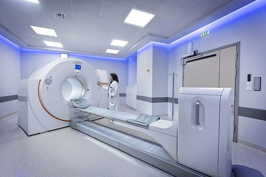
By applying two pioneering software programs that enhance the latest-generation digital PET/CT system of the Theranostics Nuclear Medicine Center at Henry Dunant, imaging results of maximum precision, quality, and reliability are now ensured. These are the FlowMotion AI and OncoFreeze AI applications, used in selected leading oncology centers in Western Europe and the United States, which mark the future of Nuclear Medicine. Henry Dunant’s new digital PET/CT is the first and only in Greece—both in the private and public sector—to incorporate both software programs, with the goal of achieving optimal imaging quality for the benefit of patients.
In practice, the two applications complement each other, offering significantly improved image quality, faster testing with less radiation, and greater reliability of results. Their application in PET/CT tests reduces imaging time by up to six minutes, leading to a more accurate evaluation of radiopharmaceutical uptake in small lesions in body regions affected by respiratory motion. It is noted that in these same regions—where constant respiratory movement during conventional imaging causes blurring and indistinct lesion borders—a clear improvement is recorded, with lesion volume reduced by 31%. This means that lesions are depicted with their true size and uptake levels within the body, which supports accurate diagnosis, contributes to reliable patient monitoring, and provides a valuable tool for the correct planning of radiotherapy.
The two software programs use Artificial Intelligence (AI) technology — not in the way many of us tend to think (i.e. that it gives us a ready-made diagnosis) — but they assist us because they ‘read’ the patient’s body and respiratory motion, and they manage to provide us with a correct image, free of motion, blurred areas, and other obstacles, so that we can interpret a higher-quality and clearer picture. This image essentially reproduces the reality inside the patient’s body — that is, we see both the structures and the lesions, such as metastases, as they truly are in terms of extent, but also in terms of radiopharmaceutical uptake,” explains Ms. Evangelia Skoura, Head of PET/CT and Deputy Director at the Theranostics Nuclear Medicine Center of Henry Dunant, adding: “Small lesions located in regions affected by respiratory motion, such as in the lungs and other thoracic structures, but also in the liver, spleen, and pancreas, might not be clearly imaged or might appear to have no uptake of radiopharmaceuticals due to the blurring caused by breathing — and as a result, possible metastases or other lesions may not be properly evaluated. With the technology now in use in Henry Dunant’s PET/CT, such small lesions are recognized with unprecedented accuracy, so that the interpretation of the test is extremely reliable, without doubts and without recommendations for further tests or repeat examinations.”
It is noted that, among other things, FlowMotion AI technology provides increased and uniform image sensitivity and precise quantification, eliminating variations in sensitivity during PET/CT scanning — a problem with traditional systems. In addition, it offers personalized scanning tailored to the patient’s body type, with reduced CT radiation exposure.
The OncoFreeze AI software minimizes the impact of breathing during scanning of the chest and upper abdomen. It allows correction of problems caused by respiratory motion during PET tests, without requiring additional acquisitions and without the use of external devices — cameras or belts (which is time-consuming and uncomfortable for the patient). As a result, clearer and more stable images are obtained in areas affected by respiratory motion (such as the lungs and liver), along with exceptional accuracy in detecting tumors or other pathological findings.